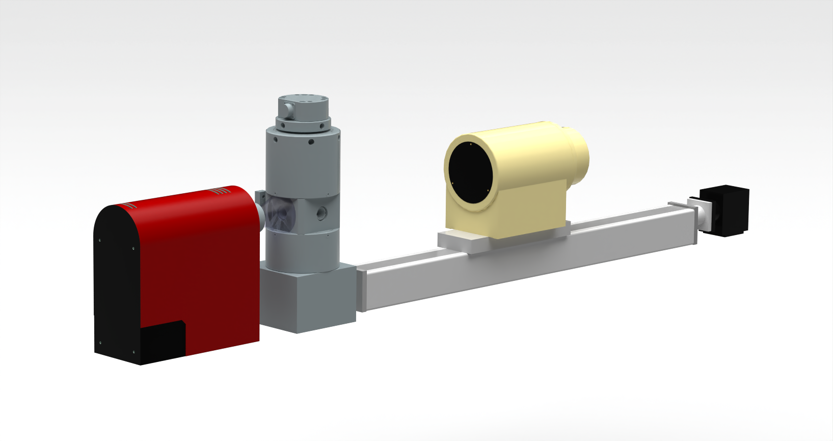
X-ray imaging (or radiography) is used to visualise the internal structure of an object, by observing the absorption contrast of its different phases. The image magnification is thus depending on the size of the source and the distance between the sample and the camera. As part of PLANEX Equipex, this custom-made experiment has been coupled to an autoclave with transparent windows. Therefore, this ensemble allows in-situ measurements of sample at high pressure and high temperature.
Scientific value :
As this autoclave is capable of withstanding extreme constraints, similar to the environmental conditions of the superior earth’s crust, it is well suited in a laboratory framework for geological application. As an example, the coupling with X-ray imaging makes it ideal to study the cristal nucleation or bubble growth within different kinds of magma.
Development :
The design of this autoclave with transparent windows is based on the one used at the FAME beamline of ESRF, dedicated to X-ray absorption spectroscopy (Testemale et al., 2005 [1]), with the support of Neel Institut and the SERAS.
Experimental conditions :
Pressure : 1 – 2000 bars
Temperature : 20 – 1200 °C
Transparent windows : beryllium (Ø 5 mm)
Nature of sample : solid, liquid
Dimensions of sample : Ø 1 – 4 mm
Sample holder materials : vitreous carbon
Technical information :
X-ray source :
XRG 60 Cu & Mo (Inel) : aperture 24°
L12161-07 W (Hamamatsu) : aperture 43°
X-ray camera C7876 0.4 MP (Hamamatsu) : 72 x 54 mm2
CCD technology coupled with image intensifier
Sample to camera distance : 10 cm – 2 m
Motorised linear translation
Data treatment software : HiPic
X-ray imaging experiment is in an advanced phase of development.
Involved collaborators in this experimental development, together with ISTO :
[1]: D. Testemale, R. Argoud, O. Geaymond and J.-L. Hazemann, High pressure/high temperature cell for x-ray absorption and scattering techniques, Review of Scientific Instruments, 2005, 76, DOI:10.1063/1.1884188